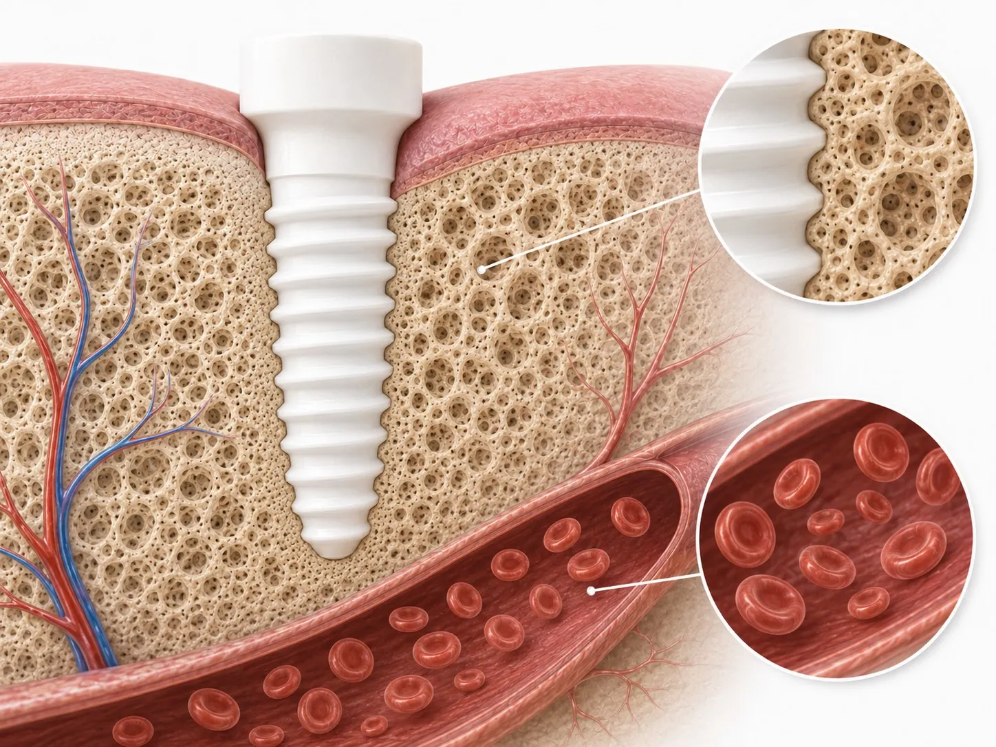
- A jawbone cavitation is an area of bone that did not fill in properly after a tooth was removed, most often at an old wisdom tooth site.
- The research is limited and debated. Some recent work suggests many of these areas may be normal variations in how sockets heal.
- That is why a decision should come from 3D imaging read together with your history, symptoms and examination, never from a scan alone.
- When surgery is chosen, it involves opening the site, removing poorly healed tissue, disinfecting the bone and supporting healing.
What a jawbone cavitation is
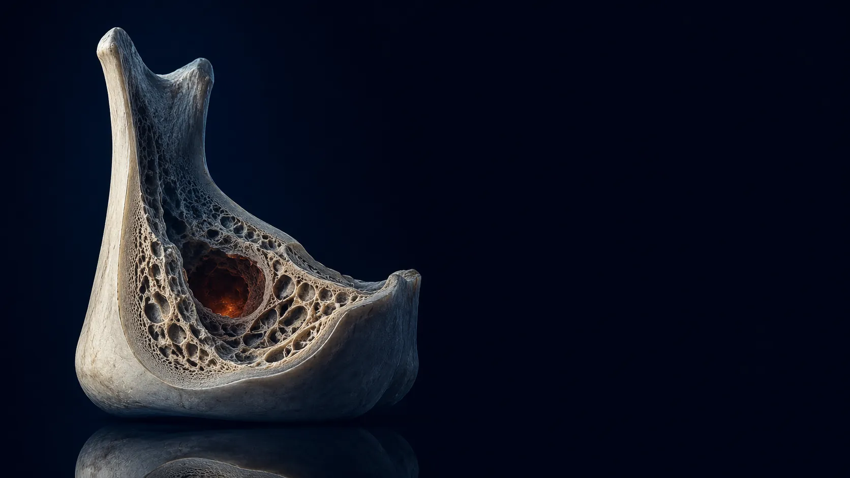
When a tooth is taken out, the empty socket is supposed to fill with new bone over the following months. Usually it does. Sometimes, particularly at old wisdom tooth sites, the gum closes over the top while the bone underneath does not fully fill in, leaving an area of poorly healed or hollow bone. Biological dentists call this a cavitation.
You will see other names in the research. NICO (neuralgia inducing cavitational osteonecrosis) was used for cases linked with facial pain, and FDOJ (fatty degenerative osteonecrosis of the jaw) is a term used by some European authors. They describe overlapping ideas, and the terminology itself is part of the debate.
An honest look at the evidence
Cavitations are one of the most discussed topics in biological dentistry, and also one of the least settled. If you are considering surgery, you deserve to know that.
- A 2022 systematic review in Oral Diseases found that the published studies on NICO were observational and of poor quality, and that the evidence on causes, diagnosis and treatment was weak.
- A recent review of the field proposes that many low density areas seen at old extraction sites on 3D scans may be a normal result of how sockets collapse and heal, and suggests a separate term for them.
- A 2025 perspective in the Journal of Pain Research questioned whether these cavities should be treated as a disease at all.
None of this means nobody ever has a problem at an old extraction site. It means a finding on a scan is a starting point for a conversation, not a diagnosis by itself.
How cavitations are found
A standard dental x-ray compresses the jaw into one flat image, so changes inside the bone can be hidden by the bone around them. Cone beam CT (CBCT) shows the jaw in three dimensions, slice by slice, which makes it far more useful for looking at old extraction sites.
Even so, imaging has limits, and not every dark area on a scan is a cavitation. A careful evaluation puts the scan together with:
- Which teeth were removed, and when
- Any pain, tenderness or other symptoms in that area, and how long they have lasted
- Your wider health history and what your physician has already ruled out
- An examination of the area itself
What cavitation surgery involves
When surgery is recommended, the aim is to clean out an area of poorly healed bone and give the site a better chance to heal. In Dr. Panahpour's practice the procedure follows five stages:
- Imaging. Cone beam CT to plan the exact location and its relationship to nerves and sinuses.
- Access. A small, minimally invasive opening at the old extraction site.
- Cleaning. Poorly healed tissue is removed and the bone is disinfected with a laser.
- Ozone. Medical grade ozone is applied to disinfect the site before it is closed.
- PRF. Platelet rich fibrin, prepared from a small sample of your own blood, is placed to support healing.
What recovery is like
Recovery is usually compared with other minor oral surgery, such as a wisdom tooth extraction, and depends on how many sites are treated and how large they are. Expect some swelling and tenderness for a few days, soft foods for a while, and specific aftercare instructions. Follow up imaging is often used later to see how the bone has filled in.
Can cavitations heal without surgery?
Not every finding needs treatment. Some areas seen on imaging may simply reflect normal healing, and some are best monitored with follow up imaging over time. Where treatment is recommended for a true area of poorly healed bone, it is generally surgical. A good clinician will tell you which category you are in, and why.
Questions to ask before cavitation surgery
- What exactly do you see on my scan, and how confident are you that it is a problem?
- What happens if we wait and image it again later?
- How do my symptoms and history fit with what you see?
- What does the surgery involve, and what are the risks for this particular site?
- If the missing tooth needs replacing later, what are my options?
Cavitation evaluation in Santa Monica and Sacramento
Dr. Panahpour evaluates old extraction sites with cone beam imaging at both offices, and reads the images together with your history and what your physician has found. Some sites are best treated and some are best watched, and you will hear both possibilities, with the reasons, before anything is scheduled.
Common questions
How do you know if you need cavitation surgery?
Only through an evaluation that combines 3D cone beam imaging with your history, your symptoms and an examination. A scan finding alone is not enough, because some low density areas at old extraction sites may be normal healing.
How long does it take to recover from cavitation surgery?
Recovery is usually compared with other minor oral surgery, such as a wisdom tooth extraction, with swelling and tenderness for a few days. It depends on the number and size of the sites treated, and you will receive specific aftercare instructions.
Can cavitations heal without surgery?
Some findings need no treatment and some are best monitored with follow up imaging. Where treatment is recommended for a true area of poorly healed bone, it is generally surgical.
What are the risks of cavitation surgery?
Like any oral surgery, risks include pain, swelling, bleeding and infection, and in some areas proximity to nerves or sinuses. 3D planning helps reduce these risks, and your dentist should explain the specific risks for your site.
Does a regular x-ray show cavitations?
Often not. A standard x-ray flattens the jaw into a single image, so changes inside the bone can be hidden. Cone beam CT shows the area in three dimensions.
Sources
- Sekundo C, et al. Neuralgia-inducing cavitational osteonecrosis: a systematic review. Oral Diseases. 2022;28(6):1448-1467
- Jawbone cavitations: current understanding and conceptual introduction of covered socket residuum (CSR). PubMed Central
- Keith DA. Neuralgia inducing cavitational osteonecrosis of the jaw: scientific controversy or pseudoscience? Journal of Pain Research. 2025;18:4275-4284

Holistic and biological dentistry since 1994, in Santa Monica and Sacramento. IAOMT accredited and SMART certified. Fourth generation dentist and author of The Good Dentist.
Related: Cavitation Surgery in Santa Monica · Zirconia Implants in Santa Monica · Oral Systemic Health in Santa Monica
This article is general education, not medical or dental advice. Every mouth and every health history is different; please talk to a licensed professional about your own situation.